Language

 动物造模,药效评价,光损伤视网膜
 z-bio
z-bio
 2024-07-18
 413
413
At present, it is believed that retinal damage caused by strong light or continuous light exposure is influenced by species, region, diet, pre-treatment of light feeding history, and genetic background. One of the mechanisms of retinal oxidative light damage is that the response of retinal oxidative damage and receptor cell damage is the result of a combination of transcriptional regulation, changes and activation of enzyme activity, intracellular metal ions, multiple apoptotic pathways, and may be species dependent.
In terms of lighting mode and intensity selection, Esfandiari et al. confirmed that alternating light and dark feeding is more in line with the daily survival status of organisms and can reduce the threshold of retinal damage in rats, resulting in more severe damage. Select adult rats aged 8-9 weeks and place them in a light dark alternating environment for adaptive feeding for 1 week. Before modeling, completely darken the rats for 12 hours and irradiate them with (5000 ± 300) lux intensity light for 3 hours, which can cause severe retinal damage in rats. According to the activity characteristics of rats, the modeling time was selected from 7:00 pm to 7:00 am the next day.
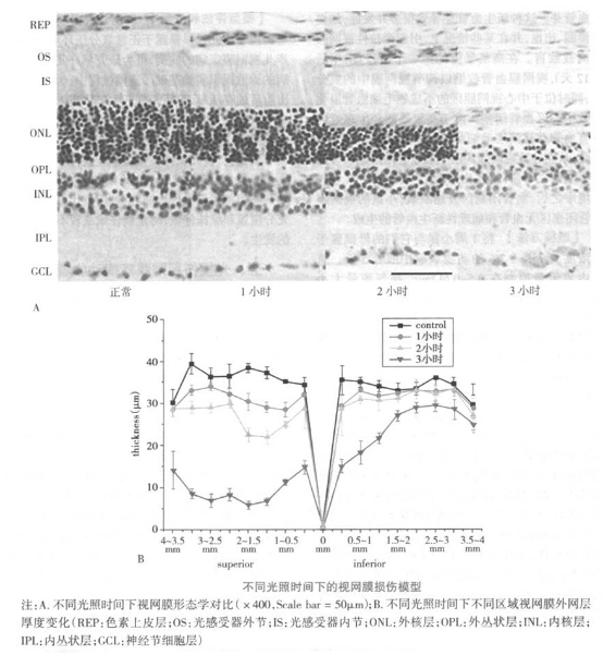
【 Model Characteristics 】 Exposure to light with an intensity of (5000 ± 300) lux for 1-3 hours can lead to a decrease in retinal electrophysiological function. The changes in various measurement indicators of ERG after different times of light irradiation are related to the length of irradiation time. The longer the irradiation time, the more obvious the changes and the more severe the damage. There is a significant statistical significance between the experimental groups. The structure of the retinal tissue slices in the normal control group rats is clear and organized, with the outer segment of the photoreceptor disk arranged neatly and regularly, and the inner and outer nuclear layers arranged tightly and stained uniformly. Strong light irradiation has a significant damaging effect on the retina, mainly located in the upper temporal quadrant of rats. Light damage is a graded response, and the degree of damage varies greatly in different regions. At different time points after light irradiation, the thickness of various regions of the retinal ONL showed significant differences.
[Model Evaluation and Application] Animal models of retinal diseases caused by light damage, due to different modeling equipment, produce varying effects. In the experiment, it is necessary to ensure that the retina of the experimental animals can be fully illuminated by light; In the selection of light intensity, the appropriate light intensity should be chosen based on the experimental purpose; When modeling animals, attention should be paid to selecting adult rats. Joly et al. reported that providing juvenile (2 or 4 weeks) and adult (8 weeks) rats with the same light intensity resulted in less light damage to the young rats, but this advantage gradually disappeared after 4 weeks; And the temperature of the modeling environment is also an important factor affecting the experimental results. Our laboratory found that high temperatures can exacerbate retinal damage.