
Supported by the National Natural Science Foundation of China (Grant No.
32150015) and other grants, the team led by Zhang Yongdeng from the School of
Life Sciences at Xihu University, in collaboration with Huang Xiaoshuai's team
from Peking University, Fan Junchao's team from Chongqing University of Posts
and Telecommunications, and Chen Liangyi's team from Peking University, has
achieved breakthroughs in the development of rapid 3D super-resolution imaging
technology for live cells. The related achievement, titled "Elucidating
Subcellular Architecture and Dynamics at Isotropic 100 nm Resolution with 4Pi
SIM Using 4Pi SIM Super Resolution Microscopic Imaging System," was published
online on December 23, 2024 in the journal Nature Methods. Paper link:
https://www.nature.com/articles/s41592-024-02515-z .
As the basic unit of life, cells have numerous organelles with different
fine structures and division of labor, performing different life activities.
Therefore, observing and studying the dynamic behavior of subcellular structures
of living cells in three-dimensional space is of great significance for the
development of cell biology. The 3D structured light illumination microscope
(3D-SIM) can increase the 3D spatial resolution to twice that of a wide field
microscope, and has the advantages of low phototoxicity, fast imaging speed, and
compatibility with traditional fluorescent probes, making it particularly
suitable for 3D delayed imaging of living cells. However, the axial resolution
of 3D-SIM (about 300 nanometers) is much lower than the lateral resolution
(about 100 nanometers), and this anisotropic resolution can lead to blurry image
details and overall distortion.
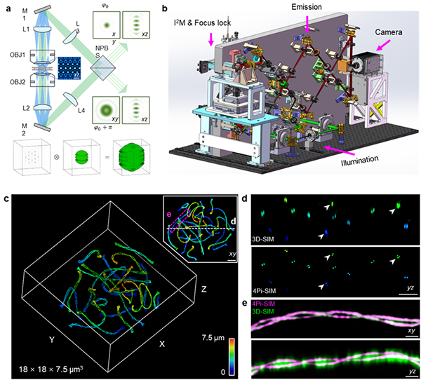
In this project, based on the research experience of 4Pi single-molecule
super-resolution microscope and Heisenberg structured light illumination
microscope, the researchers carefully designed the optical and mechanical
structures of 4Pi SIM microscope (as shown in the figure), minimizing the
influence of thermal fluctuations and mechanical vibrations, and ensuring the
long-term stability of six beam interference alignment and fluorescence
interference; By introducing a focus locking module, the system can ensure
precise alignment of two objective lenses; By designing an optical path
difference adjustment module, the system can quickly and accurately fine tune
the optical path difference between the upper and lower interference arms to
zero; In addition, the system adopts a novel lighting module and improves the
reconstruction algorithm to adaptively estimate and compensate for phase errors
caused by mismatched optical path differences in the original data, thereby
minimizing reconstruction artifacts during long-term imaging. These
technological innovations enable the 4Pi SIM system to reveal various
subcellular structures, including microtubules, endoplasmic reticulum,
mitochondria, Golgi, etc., with extremely high clarity and detail.
In addition, 4Pi SIM has achieved three-dimensional isotropic 100 nanometer
dynamic imaging on living cells for the first time, with an imaging time of
several hours, capable of capturing hundreds of time points, and a maximum
imaging speed of 0.7 Hz. Finally, 4Pi SIM also has the capability of
simultaneous dual color imaging, which can capture the rapid interaction process
between different organelles in three-dimensional space. In summary, 4Pi SIM
demonstrates the true high fidelity three-dimensional isotropic resolution
super-resolution imaging capability of live cells, taking an important step in
the field of live cell super-resolution imaging and having great potential in
elucidating subcellular dynamic behavior at the nanoscale.